-
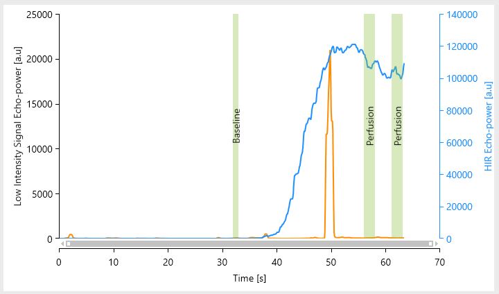
Tissue Perfusion
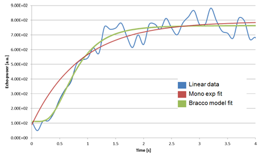
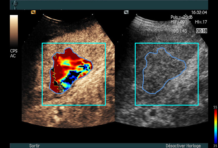
- To quantify blood perfusion in organs (non-cardiac indications).
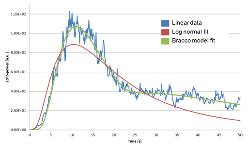
Provide a reliable method for quantifying organ perfusion, obtained from DCE-US examinations. - With specific tools to monitor perfusion of the same tissue over time.
Provide means for displaying temporal evolution of perfusion parameters on the same tissue after successive acquisitions.
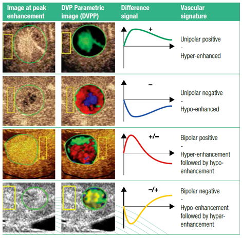
Tissue Normalization
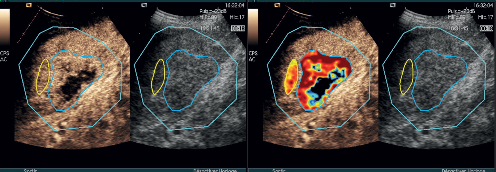
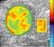
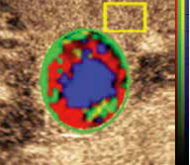
- To highlight differences in contrast enhancement patterns in different regions of interest of the analysed tissue.
Provide tools to summarize the specificities of the difference signals signatures into a single, easy-to-read color-coded image.
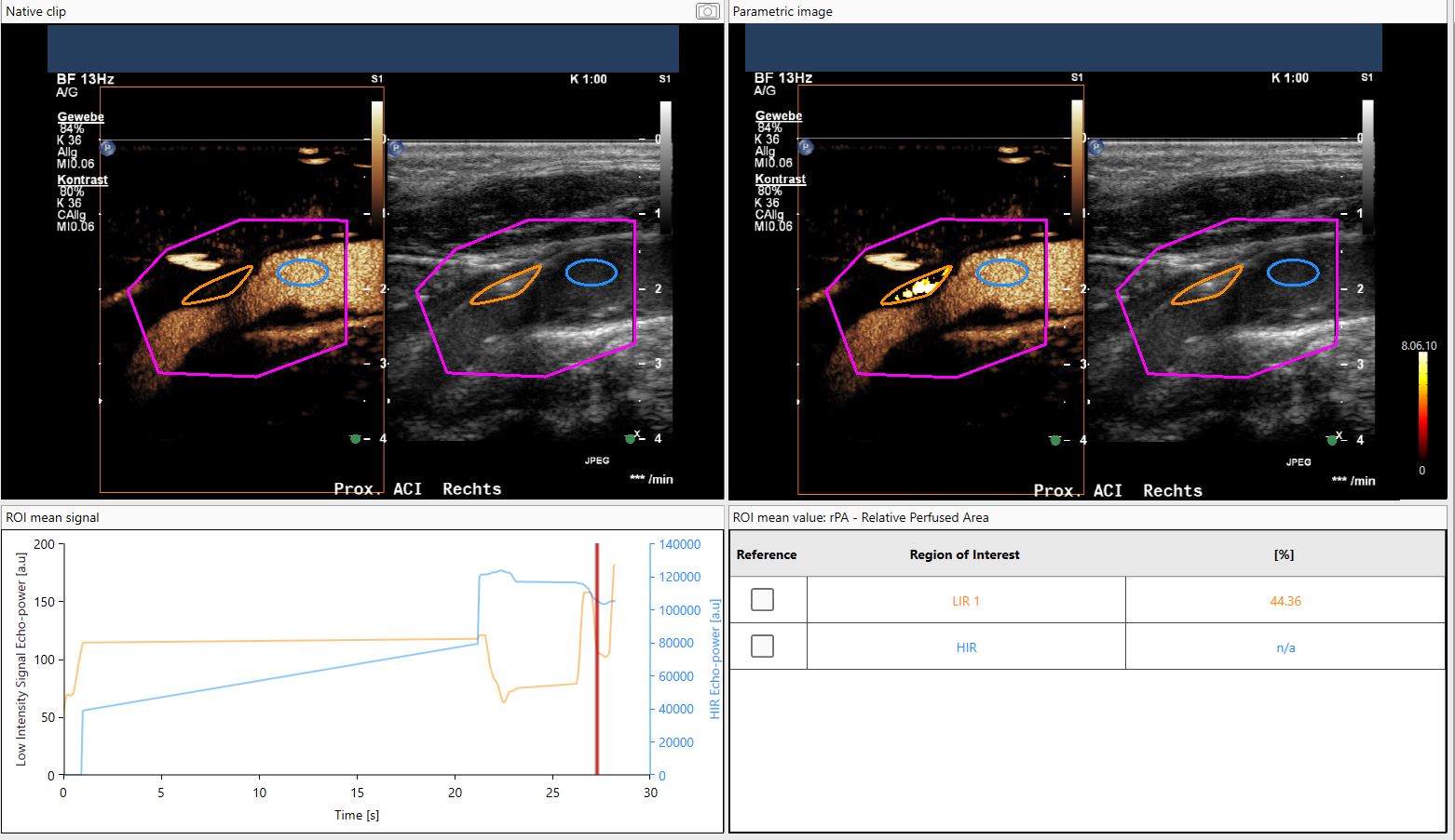
Low-Intensity Signal
- To detect vascularization and determine vascularized areas in low-intensity signal areas.
- Specific vascularization parameters: ROI area, perfused area, relative perfused area, mean opacification.
- To quantify blood perfusion in organs (non-cardiac indications).
For Research Use Only - Not for clinical diagnosis
Vuebox® Research is for RESEARCH USE ONLY and is not intended for use in diagnostic and/or clinical procedures.
For Research Use Only - Not for clinical diagnosis
